Originally a basic research tool for materials science, transmission electron microscopes (TEMs) have seen a renaissance, now being applied in nearly every technology-based field, becoming the gold standard of high spatial resolution techniques. Subsequently, these exponentially increasing applications demand a wider range of capabilities, from visualizing quantum effects to cellular 3D tomography. TEMs are used to connect photonics, nanodevice architecture, and biophysics, each with their individual intrinsic response times on the nanoscale.
Historically, “ultrafast” timescales have been driven by the available laser pulse lengths for fundamental atom-photon interactions, therefore ultrafast electron microscopy relied on the laser performance. Initially ‘ultrafast’ referred to picosecond (10-12 s, ps) time scales but has been extended through the femtosecond (10-15 s, fs) and then into the attosecond (10-18 s, as) regime with the development of laser technologies for materials processing and military applications. As these timescales are very short, it is impossible to achieve good image resolution with standard commercial electron microscopes, and therefore stroboscopic electron microscopy is required. The stroboscopic technique is easily understood by anyone that has been on a roller coaster and had their picture taken. Typically, there is a very bright flash of light to capture a decent photographic image when the object is moving at high speeds. This is because the camera’s open shutter time is very short to obtain a sharp image. The only other way to get a sharp image using the same shutter speed is to add together multiple images at lower light levels. However, these images would have to be taken at exactly the same time from the start of the ride, on many identical rides to build up a recognizable image. No closed/open eyes, no arms up/down. Exactly the same ride. Many, Many times.
Time-resolved imaging usually employs multiple pump/probe cycles because the researcher wants to know how a sample responds after a stimulus. This is typically referred to as the pump (stimulus) / probe (electron-based imaging) technique. Since images are created through stroboscopic techniques, the process being studied must be reversible so that the same image is created over many pump/probe cycles.
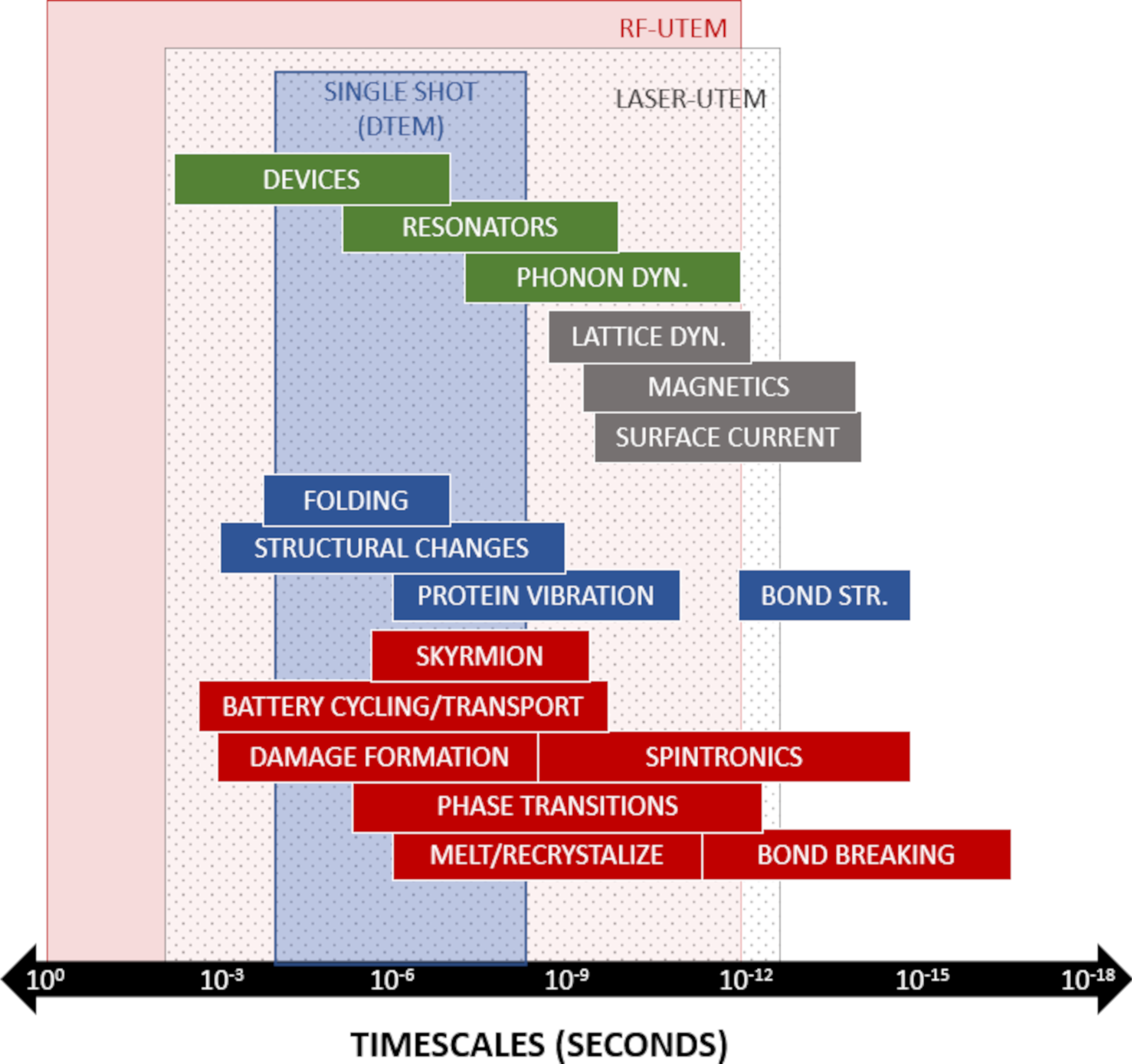
In stroboscopic ultrafast transmission electron microscopy (UTEM or more generally, UEM), the picosecond regime is common for interrogating basic material phenomena, then longer time scales are generally necessary as material systems get larger physically. The pump/probe Figure 1 shows scales of interest for time-dependent areas of study in materials science (red), life sciences (blue), semiconductor (grey) and nanotechnology (green).

Superimposed on the areas of study in Figure 1 are the typical temporal ranges for UEM techniques being discussed here. While significant temporal overlap between the UEM techniques is clear, these technologies are highly complementary to one another. Additionally, as short pulses of electrons are inherent to the UEM system, electron dose-controlled imaging can be done to reduce sample damage. Low-dose imaging isn’t usually done in a stroboscopic mode, as there is no sample stimulus except the electron beam used for the imaging.
Until recently, UTEM was possible only through laser-based methods.1,2 Laser-free UTEM has been developed, alleviating the need for laser systems with radiofrequency (RF) modulation of the TEM’s source beam.3 High frequency (GHz), sub-picosecond beam pulses have been demonstrated using a resonant deflecting cavity4,5 and a traveling wave stripline6 designs. Both techniques require no modification to the electron gun and can preserve the peak brightness and energy spread to the native source when a two-kicker design is employed. This is important because (1) user’s select TEM guns based on their research requirements, and (2) replacing the electron source with a photocathode system (as required for laser-UTEM) typically provides a beam with compromised quality, unless heroic measures are taken.7,8
- Arbouet, A., et al. (2018). Ultrafast Transmission Electron Microscopy: Historical Development, Instrumentation, and Applications. In P. W. Hawkes (ed.), Advances in Imaging and Electron Physics Volume 207 (pp. 1-72). Academic Press.
- Plemmons, D.A. and Flannigan, D.J. (2017). “Ultrafast electron microscopy: Instrument response from the single-electron to high bunch-charge regimes.” Chem Phys Lett 683(186): 186-192.
- Montgomery, E., et al. (2021). “Ultrafast Transmission Electron Microscopy: Techniques and Applications.” Microscopy Today 29(5), 46-54. doi:10.1017/S1551929521001140
- Qiu, J., et al. (2016). “GHz laser-free time-resolved transmission electron microscopy: A stroboscopic high-duty-cycle method.” Ultramicroscopy 161: 130-136. http://dx.doi.org/10.1016/j.ultramic.2015.11.006
- Verhoeven, W., et al. (2018). “High quality ultrafast transmission electron microscopy using resonant microwave cavities.” Ultramicroscopy 188 85-89. https://doi.org/10.1016/j.ultramic.2018.03.012
- Jing, C., et al. (2019). “Tunable electron beam pulser for picoseconds stroboscopic microscopy in transmission electron microscopes.” Ultramicroscopy 207: 112829. https://doi.org/10.1016/j.ultramic.2019.112829
- Feist, A., et al., (2017). “Ultrafast transmission electron microscopy using a laser-driven field emitter: Femtosecond resolution with a high coherence beam.” Ultramicroscopy 176 63-73. http://dx.doi.org/10.1016/j.ultramic.2016.12.005
- Back, N., et al., (2019). “Coulomb interactions in high-coherence femtosecond electron pulses from tip emitters.” Struct. Dyn. 6 014301. doi: 10.1063/1.5066093
